

Credit: Junxiang Zhao
A device created by electrical engineers at the University of California, San Diego, enhances the resolution of an ordinary light microscope, allowing it to directly view finer features and details in living cells.
A standard light microscope is transformed into a super-resolution microscope using this technology. It includes a specially developed substance that shortens the wavelength of light when it illuminates the sample, allowing the microscope to picture in better resolution.
Ordinary light-sheet microscopes may now detect subcellular features thanks to a hyperbolic metamaterial developed at the University of California, San Diego. In other words, an ordinary light microscope can now see in superresolution. As the sample is irradiated, the material, which is made up of nanometer-thin alternating layers of silica glass and silver, shortens the wavelength of light, allowing the microscope to see below the diffraction limit.
According to a professor of electrical and computer engineering at UC San Diego, Zhaowei Liu the light-shrinking material turns low-resolution light into high-resolution light in the application. Liu explained that It’s incredibly basic and straightforward to use. All you have to do is put a sample on the material and then put the whole thing under a standard microscope.
The research solves a significant flaw in conventional light microscopes, as well as a flaw in superresolution imaging approaches in general. Light microscopes are beneficial for observing live cells, and other techniques, such as electron microscopy, enable the analyst to see subcellular structures, though the sample must be placed in a vacuum chamber. Other methods, such as expansion microscopy, involve changing the cells. Although conventional light microscopes with a resolution of 200 nm are unable to examine anything closer than that distance as separate objects, light microscopy allows researchers to examine living cells in their natural condition.

Credit: Junxiang Zhao
Drawback Of Original Light Microscopes
The research, which was reported in Nature Communications, addresses a major drawback of original light microscopes: low resolution. Light microscopes are great for viewing live cells, but they can’t see anything smaller than that. The resolution limit of conventional light microscopes is 200 nanometers, which means that things closer than this distance will not be seen as independent objects.
The main problem is to create a technology that has extremely high resolution while also being suitable for live cells,” Liu explained.
Both characteristics are combined in the technology created by Liu’s team. With it, a conventional light microscope can be utilized to picture live subcellular structures with a 40 nanometers resolution.
A microscope slide coated with a hyperbolic metamaterial, a sort of light-shrinking material, is used in the technology. It is made up of silica glass layers and nanometer-thin silver that alternate. Light’s wavelengths scatter and shorten as it passes through, resulting in a series of random high-resolution speckled patterns. When a sample is mounted on the slide, this series of speckled light patterns illuminate it in various ways. This results in a series of low-resolution images being collected, which are then stitched together using a reconstruction algorithm to generate a high-resolution image.
Credit: Nature Communications
Application of Ordinary Microscope
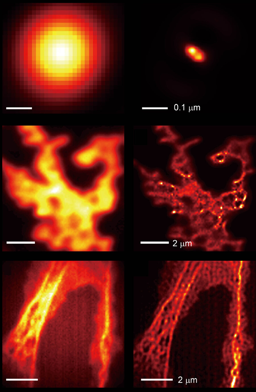
A commercial inverted microscope was used to test the researchers’ technology. They were able to view small details in fluorescently labeled Cos-7 cells — features, like actin filaments, that would be difficult to see with merely a microscope. The researchers were also able to discriminate between tiny fluorescent beads and quantum dots spaced 40 to 80 nanometers apart thanks to the technique.
According to the researchers, super-resolution innovation offers a lot of potential for high-speed operation. Their goal for live-cell imaging is to combine high speed, super-resolution, and minimal phototoxicity in a single system.

Credit: Yeon Ui Lee
Liu’s team is now working to improve the technology so that it can undertake high-resolution imaging in three dimensions. Liu and his colleagues earlier published a paper demonstrating that this method can also image with ultra-high axial resolution (about 2 nanometers). They’re currently working on integrating the two.
Sources:
Main research paper: Yeon Ui Lee et al, Metamaterial assisted illumination nanoscopy via random super-resolution speckles, Nature Communications (2021). DOI: 10.1038/s41467-021-21835-8
https://phys.org/news/2021-06-light-shrinking-material-ordinary-microscope-super.html
https://www.photonics.com/Articles/Metamaterial_Lets_Light-Sheet_Microscopes_See_in/a67044


